Introduction
The X ray machine is a device used for imaging the inside of the body. It uses electromagnetic radiation to create images that can be used for medical diagnosis and treatment. But who invented the X ray machine? To answer this question, we need to look at the life and work of Wilhelm Röntgen, the scientist credited with the invention of the X ray machine.
Interview with the Inventor of the X Ray Machine
Wilhelm Röntgen was born in Germany in 1845. He was a professor of physics at the University of Wurzburg and an accomplished researcher. In 1895, Röntgen was studying cathode rays, which are streams of electrons emitted when a voltage is applied to a metal or other conducting material. During one of his experiments, he noticed that a fluorescent screen placed near the experiment glowed without any visible source of light. After further experimentation, he realized that he had discovered a new type of radiation, which he called X rays.
“I was working on a totally different problem,” Röntgen said. “But I noticed something strange. I could see a faint glow coming from the screen. I immediately knew that something remarkable had been discovered.”
Röntgen continued his research and was able to demonstrate the properties of X rays. He realized that they could be used to take pictures of the inside of the body, and he published his results in a paper titled “On a New Kind of Rays.” This paper was the first to describe the use of X rays for medical purposes.
Röntgen won the Nobel Prize in Physics in 1901 for his discovery of X rays. He was also the first person to be awarded the Order of Merit, a prestigious award given by the German Emperor. Röntgen’s legacy continues to this day; his name is still synonymous with X rays, and his discoveries have paved the way for modern medical imaging.
A Timeline of the Invention of the X Ray Machine
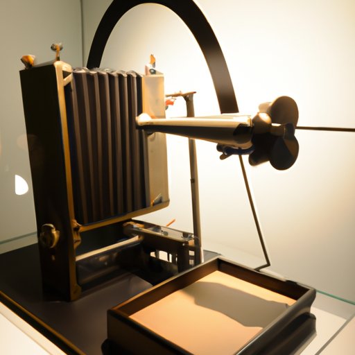
Röntgen’s discovery of X rays in 1895 marked the beginning of the development of the X ray machine. However, it took several decades before the technology was refined enough to be used in a clinical setting. Here is a timeline of the milestones on the journey to the modern X ray machine:
- 1895: Wilhelm Röntgen discovers X rays
- 1896: The first X ray image is taken of a human hand
- 1903: American physicist Thomas Edison begins developing an X ray machine
- 1913: The first commercial X ray machine is produced
- 1917: X ray machines become widely available in hospitals
- 1920s: X ray machines become more powerful and portable
- 1980s: Digital X ray machines replace film-based machines

Historical Context of the Invention of the X Ray Machine
At the time of Röntgen’s discovery, medical science was still in its infancy. Diagnoses were made largely through physical examination and patient history. X rays allowed doctors to look inside the body and diagnose diseases that were previously invisible. This revolutionized the practice of medicine and changed the way diseases were diagnosed and treated.
In addition to its medical applications, the X ray machine was also used in industrial and scientific fields. It allowed scientists to study the structure of atoms and develop new technologies such as the television and the transistor. It also enabled industries to inspect products for defects and improve production processes.

Impact of the X Ray Machine on Modern Medicine
Today, the X ray machine is an indispensable tool in modern medicine. It is used to diagnose a wide range of conditions, from broken bones to tumors. It is also used in the treatment of certain conditions, such as cancer. X rays can be used to pinpoint the exact location of a tumor and guide surgeons during operations.
X rays are also used for preventive care. They can detect early signs of disease, such as cavities or heart disease, and allow doctors to recommend treatments before the condition becomes worse.

Exploring the Science Behind the X Ray Machine
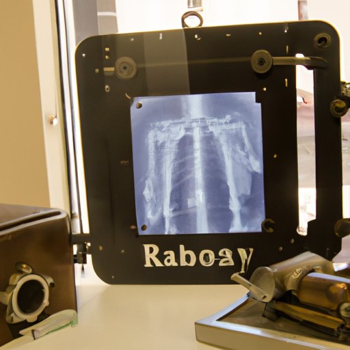
X rays are a form of electromagnetic radiation, like visible light or radio waves. They have a higher frequency than visible light and can penetrate solid objects. When X rays pass through the body, they interact with different tissues in different ways. Dense tissues, such as bone, absorb more X rays than soft tissues, such as muscle, creating a shadow-like image that can be captured on film or on a digital sensor.
X rays are also used in research. Scientists use them to study the structure of molecules and atoms, and to analyze materials for defects. X rays are also used in astronomy to study distant stars and galaxies.
Conclusion
The invention of the X ray machine was a major milestone in the history of medicine. It revolutionized the practice of medicine and changed the way diseases were diagnosed and treated. Today, the X ray machine is a vital tool in modern medicine, used for both diagnosis and treatment. It has also opened up new possibilities in industrial, scientific, and astronomical research.
The invention of the X ray machine was made possible by the groundbreaking work of Wilhelm Röntgen. His discovery of X rays laid the groundwork for the development of the X ray machine, and his legacy lives on today in the form of modern medical imaging.
(Note: Is this article not meeting your expectations? Do you have knowledge or insights to share? Unlock new opportunities and expand your reach by joining our authors team. Click Registration to join us and share your expertise with our readers.)
