Introduction
Fundus photography is a specialized imaging technique used to capture detailed images of the retina and other parts of the eye. It is a valuable tool for diagnosing and monitoring various eye diseases, such as macular degeneration, glaucoma, and diabetic retinopathy. In this article, we will explore what fundus photography is and how it works, its benefits in eye care, the types of cameras used, the process of performing a fundus photographic examination, and its different uses and applications in ophthalmology.
What is Fundus Photography?
Fundus photography is a specialized imaging technique used to capture detailed images of the retina, optic disc, macula, and other parts of the eye. It is also known as ophthalmoscopy or retinal photography. The purpose of this imaging technique is to document the appearance of the back of the eye and detect any abnormalities or changes that may indicate a disease or condition. Fundus photography has been used in ophthalmology since the early 1900s and is now an integral part of routine eye exams.

Overview of How It Works
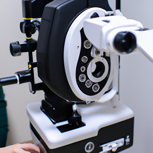
Fundus photography uses a special camera called a fundus camera to take pictures of the back of the eye. A fundus camera consists of a light source, a lens, and a digital camera. The light source illuminates the back of the eye and the lens magnifies the image so that it can be captured by the digital camera. The resulting images are then stored digitally and can be viewed on a computer monitor or printed out for further review.

Benefits of Fundus Photography in Eye Care
Fundus photography offers numerous benefits in eye care, including improved diagnostic accuracy, early detection of disease, and increased treatment options. According to a study published in the journal Ophthalmic and Physiological Optics, “Fundus photography is an invaluable tool for documenting retinal pathology and aiding in the diagnosis and management of many ocular conditions.”
Improved Diagnostic Accuracy
Fundus photography allows doctors to get a more detailed and accurate view of the back of the eye than traditional methods, such as direct ophthalmoscopy. This can help them diagnose diseases and conditions more precisely, leading to better outcomes for patients.
Early Detection of Disease
Fundus photography can also help detect diseases at an earlier stage, when they may be more easily treatable. For example, it can be used to detect signs of macular degeneration before it causes permanent vision loss.
Increased Treatment Options
Fundus photography can also provide doctors with additional information to aid in their decision-making process when selecting treatments for certain eye conditions. For instance, it can help them determine whether a patient would benefit from laser surgery or medications.
Examining the Fundus Camera
When selecting a fundus camera, there are several factors to consider. Different types of cameras are available, and each one has its own advantages and disadvantages. Additionally, there are several components of a fundus camera that must be taken into account, such as the type of illumination, the size and resolution of the digital camera, and the magnification of the lens.
Types of Cameras Used
The three main types of fundus cameras are slit-lamp fundus cameras, direct fundus cameras, and indirect fundus cameras. Slit-lamp fundus cameras are typically used for examining small areas of the retina, such as the macula. Direct fundus cameras allow for a wider field of view and are often used for general examinations of the retina. Indirect fundus cameras use a mirror to reflect the image onto the camera’s sensor, allowing for a larger field of view.
Components of a Fundus Camera
The components of a fundus camera include a light source, a lens, and a digital camera. The light source is used to illuminate the back of the eye and the lens magnifies the image so that it can be captured by the digital camera. The size and resolution of the digital camera will affect the quality of the images produced.
Factors to Consider When Choosing a Fundus Camera
When choosing a fundus camera, it is important to consider the type of camera, the size and resolution of the digital camera, the amount of magnification provided by the lens, and the type of illumination used. Additionally, cost is an important factor to consider when selecting a fundus camera.
The Process of Performing a Fundus Photographic Examination
Performing a fundus photographic examination requires careful preparation and a series of steps. First, the patient must be properly positioned and prepared for the examination. Then, the eye must be dilated in order to achieve the best possible image quality. The fundus camera must then be properly aligned and focused on the eye. Finally, the images are captured and stored digitally for further review.

Understanding the Uses and Applications of Fundus Photography in Ophthalmology
Fundus photography is an invaluable tool for diagnosing and monitoring various eye diseases. It can be used to detect diseases at an earlier stage, improve diagnostic accuracy, and increase treatment options. Additionally, it can produce different types of images, such as color images, fluorescein angiograms, and autofluorescence images. However, there are some potential issues with fundus photography, such as difficulty focusing the camera, poor image quality, and artifacts created by the camera.
Conclusion
Fundus photography is an important imaging technique used to capture detailed images of the retina and other parts of the eye. It offers numerous benefits in eye care, including improved diagnostic accuracy, early detection of disease, and increased treatment options. Additionally, it can be used to produce different types of images, such as color images and fluorescein angiograms. Fundus photography is an invaluable tool for diagnosing and monitoring various eye diseases and should be a part of every comprehensive eye exam.
(Note: Is this article not meeting your expectations? Do you have knowledge or insights to share? Unlock new opportunities and expand your reach by joining our authors team. Click Registration to join us and share your expertise with our readers.)
