Introduction
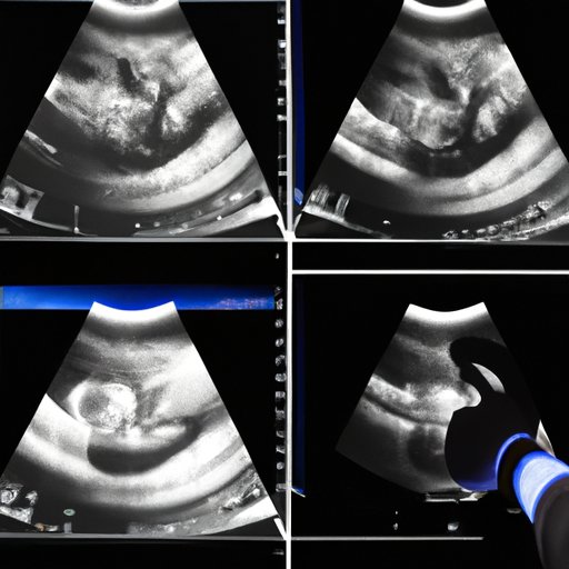
Ultrasound imaging is a widely used medical imaging technology that has revolutionized the diagnosis and treatment of illnesses and injuries. Ultrasound imaging uses sound waves to create an image of the inside of the body. This technology is non-invasive, meaning no needles or radiation is used. Ultrasound imaging can be used to diagnose and monitor a variety of medical conditions, from pregnancy to cancer.
The purpose of this article is to explore the different types of ultrasound imaging technology and the advantages and disadvantages of each. We will look at two-dimensional (2D) ultrasound imaging, three-dimensional (3D) ultrasound imaging, four-dimensional (4D) ultrasound imaging, Doppler imaging, contrast-enhanced ultrasound imaging, tissue harmonic imaging, and elastography.
2D Ultrasound Imaging
2D ultrasound imaging is the most common type of ultrasound imaging. It uses high-frequency sound waves to create an image of the inside of the body. The sound waves are sent into the body, then bounce off internal organs and tissues and return back to the transducer, which is a device that converts the sound signals into images. These images are displayed on a monitor.
The advantages of 2D ultrasound imaging include its affordability and availability, as well as its ability to detect certain types of diseases and abnormalities. However, 2D ultrasound imaging has some drawbacks. It is not as detailed as other types of imaging technology, and it may not be able to detect smaller abnormalities. Additionally, the accuracy of 2D ultrasound imaging can vary depending on the experience of the technician.
3D Ultrasound Imaging
3D ultrasound imaging is an advanced form of ultrasound imaging. It uses multiple sound beams to create a three-dimensional view of the inside of the body. This allows for more detail than 2D ultrasound imaging, and it can help physicians better diagnose and monitor medical conditions. 3D ultrasound imaging can also be used to measure the size, shape, and volume of organs and tissues.
The advantages of 3D ultrasound imaging include its higher level of detail, its ability to provide measurements, and its potential to improve diagnosis and treatment decisions. However, 3D ultrasound imaging is expensive and not widely available. Additionally, 3D ultrasound imaging may not always be necessary for certain types of medical procedures.
4D Ultrasound Imaging
4D ultrasound imaging is an advanced form of 3D ultrasound imaging. It uses multiple sound beams to create a three-dimensional image in real time. This allows physicians to observe how the organ or tissue moves in real time, which can help them diagnose and treat certain medical conditions. 4D ultrasound imaging is often used to monitor fetal development during pregnancy.
The advantages of 4D ultrasound imaging include its ability to provide real-time images of the organ or tissue, its potential to improve diagnosis and treatment decisions, and its potential to improve prenatal care. However, 4D ultrasound imaging is expensive and not widely available. Additionally, it is not always necessary for certain types of medical procedures.
Doppler Imaging
Doppler imaging is an advanced form of ultrasound imaging. It uses sound waves to measure the speed and direction of blood flow. This information can be used to diagnose and treat a variety of medical conditions, such as arterial blockages and heart valve problems. Doppler imaging can also be used to monitor the progress of medical treatments, such as stents or angioplasty.
The advantages of Doppler imaging include its ability to measure blood flow, its potential to improve diagnosis and treatment decisions, and its potential to monitor the progress of medical treatments. However, Doppler imaging is expensive and not widely available. Additionally, it is not always necessary for certain types of medical procedures.
Contrast-Enhanced Ultrasound Imaging
Contrast-enhanced ultrasound imaging is an advanced form of ultrasound imaging. It uses an ultrasound contrast agent, which is a liquid that contains tiny bubbles that reflect sound waves, to improve the clarity of the images. This can help physicians better diagnose and treat certain medical conditions, such as cancer or kidney stones.
The advantages of contrast-enhanced ultrasound imaging include its ability to improve image clarity, its potential to improve diagnosis and treatment decisions, and its potential to monitor the progress of medical treatments. However, contrast-enhanced ultrasound imaging is expensive and not widely available. Additionally, it is not always necessary for certain types of medical procedures.
Tissue Harmonic Imaging
Tissue harmonic imaging is an advanced form of ultrasound imaging. It uses sound waves of different frequencies to create a clearer image of the inside of the body. This technology can help physicians better diagnose and treat a variety of medical conditions, including tumors and cysts.
The advantages of tissue harmonic imaging include its ability to improve image clarity, its potential to improve diagnosis and treatment decisions, and its potential to monitor the progress of medical treatments. However, tissue harmonic imaging is expensive and not widely available. Additionally, it is not always necessary for certain types of medical procedures.
Elastography
Elastography is an advanced form of ultrasound imaging. It uses sound waves to measure the stiffness of tissues and organs. This information can be used to diagnose and treat a variety of medical conditions, such as cancer, fibroids, and liver disease. Elastography can also be used to monitor the progress of medical treatments.
The advantages of elastography include its ability to measure tissue stiffness, its potential to improve diagnosis and treatment decisions, and its potential to monitor the progress of medical treatments. However, elastography is expensive and not widely available. Additionally, it is not always necessary for certain types of medical procedures.
Conclusion
Ultrasound imaging is a powerful and versatile medical imaging technology. There are many different types of ultrasound imaging, including 2D, 3D, 4D, Doppler, contrast-enhanced, tissue harmonic, and elastography. Each type of imaging has its own advantages and disadvantages, but all of them have the potential to improve diagnosis and treatment decisions.
Overall, ultrasound imaging is a safe and effective way to diagnose and monitor a variety of medical conditions. It is also relatively affordable and widely available, making it a popular choice for healthcare providers and patients alike.
(Note: Is this article not meeting your expectations? Do you have knowledge or insights to share? Unlock new opportunities and expand your reach by joining our authors team. Click Registration to join us and share your expertise with our readers.)
