Introduction
X-rays are a form of electromagnetic radiation that can penetrate through solid objects, allowing us to see what lies beneath the surface. X-ray machines are used in medical imaging to provide detailed images of the inside of the body. In this article, we will explore how an X-ray machine works and examine the components, mechanics, and physics behind it.
Exploring the Components of an X-Ray Machine and How They Work Together
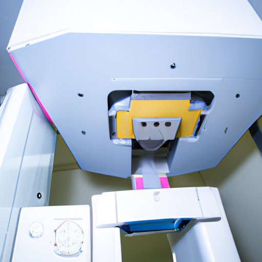
An X-ray machine consists of several components that work together to produce an X-ray image. The components include an X-ray tube, a high-voltage generator, a collimator, and a detector.
The X-ray tube is the source of the X-ray beam. It is composed of an evacuated glass envelope containing a cathode and an anode. When a high voltage is applied to the X-ray tube, electrons are emitted from the cathode and accelerated towards the anode. As the electrons strike the anode, they produce X-rays.
The high-voltage generator is responsible for supplying the X-ray tube with the necessary voltage. The collimator is used to shape the X-ray beam into a narrow beam that is focused on the area of interest. Finally, the detector captures the X-ray image and converts it into a digital signal that can be displayed on a monitor.
These components work together to produce an X-ray image. First, the high-voltage generator supplies the X-ray tube with the necessary voltage. Then, the X-ray beam is shaped by the collimator and focused on the area of interest. Finally, the detector captures the X-ray image and displays it on a monitor.
A Step-by-Step Guide to Understanding the Mechanics of an X-Ray Machine
Now that we’ve explored the components of an X-ray machine, let’s take a look at how they work together to create an X-ray image. Here is a step-by-step guide to understanding the mechanics of an X-ray machine:
Step 1: Preparing the patient for the X-ray. The first step is to prepare the patient for the X-ray. This includes positioning the patient so that the area of interest is in the center of the X-ray field. Additionally, the patient may be asked to hold their breath during the imaging process.
Step 2: Focusing the X-ray beam. Next, the X-ray beam must be focused on the area of interest. This is done by adjusting the collimator to shape the X-ray beam into a narrow beam that is focused on the area of interest.
Step 3: Generating the X-ray beam. Once the X-ray beam is focused, the X-ray tube produces the X-ray beam. This is done by applying a high voltage to the X-ray tube, which causes electrons to be emitted from the cathode and accelerated towards the anode. As the electrons strike the anode, they produce X-rays.
Step 4: Capturing the X-ray image. Finally, the X-ray image is captured by the detector and converted into a digital signal that can be displayed on a monitor. This allows the radiologist or technician to view the X-ray image and make a diagnosis.
Examining the Physics Behind an X-Ray Machine
Now that we’ve examined the mechanics of an X-ray machine, let’s take a look at the physics behind it. To understand how an X-ray machine works, it’s important to understand what an X-ray is and how it penetrates through objects.
What is an X-ray? An X-ray is a form of electromagnetic radiation that has a shorter wavelength than visible light. X-rays are able to pass through solid objects, such as the human body, and are used in medical imaging to provide detailed images of the inside of the body.
How does an X-ray machine generate an X-ray beam? An X-ray machine generates an X-ray beam by applying a high voltage to an X-ray tube. This causes electrons to be emitted from the cathode and accelerated towards the anode. As the electrons strike the anode, they produce X-rays.
How does an X-ray penetrate through objects? An X-ray is able to penetrate through objects because it has a shorter wavelength than visible light. X-rays are able to pass through soft tissue, but they are absorbed by denser materials such as bone. This allows X-rays to create detailed images of the inside of the body.
The Different Types of X-Ray Machines and Their Functions
There are several types of X-ray machines that are used for different purposes. These include stationary X-ray machines, portable X-ray machines, and digital X-ray machines.
Stationary X-ray machines: Stationary X-ray machines are typically found in hospitals and clinics. They are large and bulky, but they are capable of producing high-quality images. They are used for a variety of imaging procedures, including chest X-rays, mammograms, and dental X-rays.
Portable X-ray machines: Portable X-ray machines are smaller and more lightweight than stationary X-ray machines. They are often used in emergency rooms and ambulances, as they can be quickly moved around. They are used for imaging procedures such as chest X-rays and abdominal X-rays.
Digital X-ray machines: Digital X-ray machines use digital technology to produce X-ray images. They have the advantage of being able to store the images electronically, which makes them easier to access and share. They are often used for imaging procedures such as CT scans and MRI scans.
How X-Ray Machines Have Evolved Over Time
X-ray machines have come a long way since their invention in 1895. Early X-ray machines were bulky and required long exposure times to produce an image. Modern X-ray machines are much smaller and can produce images much faster.
Early X-ray machines used film to capture the X-ray image. This was a slow and tedious process, as the film had to be developed after each exposure. Modern X-ray machines use digital detectors to capture the X-ray image, which is then stored electronically. This makes it much easier to access and share the images.
In addition, modern X-ray machines use computer-aided detection (CAD) software to analyze the images. This software is able to detect any abnormalities in the images, which can help improve accuracy and reduce the need for additional imaging.
Conclusion
X-ray machines are an invaluable tool for medical imaging. They allow us to see what lies beneath the surface, providing detailed images of the inside of the body. In this article, we explored how an X-ray machine works and examined the components, mechanics, and physics behind it. We also looked at the different types of X-ray machines and how they have evolved over time.
X-ray machines are an essential part of modern medicine, and their importance cannot be overstated. Understanding how they work is key to unlocking their potential and improving patient care.
(Note: Is this article not meeting your expectations? Do you have knowledge or insights to share? Unlock new opportunities and expand your reach by joining our authors team. Click Registration to join us and share your expertise with our readers.)