Introduction
Magnetic resonance imaging (MRI) is a technology used to create detailed images of the body’s internal structures. MRI scans are commonly used for medical diagnosis and can provide insight into conditions such as cancer, stroke, and heart disease. In this article, we will explore how an MRI works, from the steps involved in the process to the safety of the procedure. We will also highlight the benefits of an MRI scan, including accuracy, non-invasiveness, and wide range of applications.
Explain the MRI Process Step-by-Step
The MRI process consists of several steps, beginning with the patient lying on a table that slides inside the MRI machine and ending with the radiologist interpreting the resulting images. Let’s take a closer look at each step:
Overview of the Process
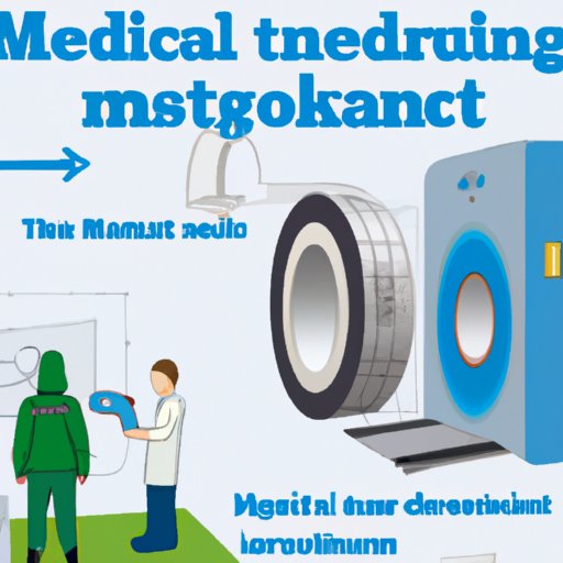
Before the MRI scan begins, the patient must first remove any metal objects they are wearing, such as jewelry or eyeglasses, as these can interfere with the MRI machine. The patient then lies on the table and is moved into the center of the magnet. During the scan, powerful magnets, radio waves, and computer systems work together to create detailed images of the patient’s body. After the scan is complete, the images are analyzed by a radiologist and a report is sent to the patient’s doctor.
In-depth Look at Each Step
1. Preparation: Before the MRI, the patient must first remove any metal items they are wearing, such as jewelry or eyeglasses, as these can interfere with the MRI machine. The patient may also need to drink a contrast agent to help improve the clarity of the images.
2. Positioning: The patient lies on the table and is moved into the center of the magnet. The patient may be asked to stay still during the scan, or may be given a sedative to help them remain still.
3. Scanning: During the scan, powerful magnets, radio waves, and computer systems work together to create detailed images of the patient’s body. The patient may hear loud noises during the scan, which can last anywhere from 15 minutes to over an hour.
4. Analysis: After the scan is complete, the images are analyzed by a radiologist and a report is sent to the patient’s doctor. The report will include information about any abnormalities found and will help the doctor diagnose and treat the patient’s condition.

Compare and Contrast MRI with Other Imaging Techniques
MRI is one of several imaging techniques used to diagnose medical conditions. Let’s compare and contrast MRI with other imaging techniques, such as X-rays, CT scans, and ultrasound:
X-Rays
X-rays use radiation to create images of the body’s internal structures. While X-rays can be used to diagnose a variety of conditions, they cannot provide detailed images like an MRI. Additionally, X-rays expose the patient to radiation, which can increase their risk of developing cancer.
CT Scans
CT scans use X-rays and a computer to create detailed images of the body’s internal structures. Like X-rays, CT scans expose the patient to radiation, but they can provide more detailed images than an X-ray. However, they cannot provide the same level of detail as an MRI.
Ultrasound
Ultrasound uses sound waves to create images of the body’s internal structures. Ultrasound is a non-invasive, painless procedure and does not expose the patient to radiation. However, it cannot provide the same level of detail as an MRI.
Describe How an MRI Machine Works
An MRI machine consists of three main components: magnetic field generators, radio frequency coils, and computer systems. Let’s take a closer look at each component:
Magnetic Field Generators
The magnetic field generators are the most important part of the MRI machine, as they generate a powerful magnetic field that aligns the protons in the patient’s body. This alignment allows the radio waves to generate signals that are used to create the images.
Radio Frequency Coils
The radio frequency coils are used to transmit and receive the radio waves. They are placed around the patient’s body and send and receive signals that are converted into images by the computer system.
Computer Systems
The computer system is responsible for analyzing the signals from the radio frequency coils and converting them into images. The images are then displayed on a monitor and can be manipulated to provide different views of the patient’s body.

Discuss the Safety of MRI Procedures
MRI scans are generally safe and pose very little risk to the patient. However, there are some risks associated with the procedure, such as claustrophobia and anxiety. Patients with implants or metal objects in their body may also be at risk for complications, as these objects can be affected by the magnetic field. Additionally, patients who are pregnant or have kidney disease should speak to their doctor before undergoing an MRI scan.
There are also some side effects associated with the procedure. These include nausea, dizziness, headache, and skin irritation. Most of these side effects are minor and go away quickly, but patients should speak to their doctor if they experience any severe or lasting side effects.

Highlight the Benefits of MRI Scans
MRI scans offer many benefits, including accuracy, non-invasiveness, and a wide range of applications. Let’s take a closer look at each benefit:
Accuracy
MRI scans are highly accurate and can provide detailed images of the body’s internal structures. According to a study published in the journal Radiology, MRI scans have a sensitivity of 97% and a specificity of 95%. This means that MRI scans are able to detect abnormalities with a high degree of accuracy.
Non-Invasive
Unlike other imaging techniques, such as CT scans and X-rays, MRI scans are non-invasive and do not expose the patient to radiation. This makes them safer than other imaging techniques and reduces the risk of complications.
Wide Range of Applications
MRI scans can be used to diagnose a wide range of medical conditions, including cancer, stroke, and heart disease. They can also be used to monitor the progression of certain conditions, as well as to assess the effectiveness of treatments.
Conclusion
In conclusion, MRI is a powerful imaging technique that can provide detailed images of the body’s internal structures. The MRI process consists of several steps, beginning with the patient lying on a table that slides inside the MRI machine and ending with the radiologist interpreting the resulting images. MRI scans are generally safe and pose very little risk to the patient, and offer many benefits, including accuracy, non-invasiveness, and a wide range of applications.
Overall, MRI is a valuable tool for diagnosing and treating a variety of medical conditions. It is important for patients to understand the process, safety, and benefits of an MRI scan so they can make an informed decision about whether or not to proceed with the procedure.
(Note: Is this article not meeting your expectations? Do you have knowledge or insights to share? Unlock new opportunities and expand your reach by joining our authors team. Click Registration to join us and share your expertise with our readers.)
