Introduction
An electrocardiogram (EKG) is a diagnostic tool used to measure electrical signals in the heart. The information gathered from an EKG can be used to diagnose cardiovascular conditions, such as arrhythmias, coronary artery disease, and heart attack. This article will explore how an EKG works, from the anatomy of the machine to the science behind the procedure. A guide to understanding the basics of an EKG, a look at the electrical signals measured by an EKG, and a step-by-step guide to performing an EKG are also included.

Exploring the Components of an EKG: An Overview of How an EKG Works
An EKG machine consists of several components that work together to measure electrical signals in the heart. These components include electrodes, wires, and a monitor.
Anatomy of an EKG Machine
The primary component of an EKG machine is the electrodes. The electrodes are placed on the skin of the patient’s chest, arms, and legs. They detect the electrical signals generated by the heart and send them to the EKG machine via wires. The EKG machine then amplifies the signals and displays them on the monitor.
The Process of Measuring Electrical Signals with an EKG
The electrodes on the patient’s skin detect small electrical signals produced by the heart. These signals are sent to the EKG machine via wires. The machine then amplifies the signals and displays them on the monitor. The monitor shows a graph of the electrical activity of the heart, which can be interpreted by a healthcare provider to diagnose any potential problems.

The Science Behind an EKG: Examining the Mechanics of the Procedure
An EKG measures the electrical signals generated by the heart. To understand how an EKG works, it is important to understand the electrical signals in the heart and how they are measured.
Electrical Signals in the Heart
The heart has four chambers, each of which contains muscle cells that contract and relax in a coordinated manner. When these muscle cells contract, they produce small electrical signals that travel through the heart. These signals are detected by the electrodes on the patient’s skin and sent to the EKG machine.
How an EKG Measures These Signals
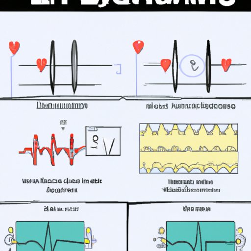
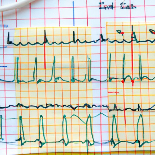
The EKG machine amplifies the signals and displays them on the monitor. The monitor shows a graph of the electrical activity of the heart, which can be interpreted by a healthcare provider to diagnose any potential problems. The graph is divided into three sections: the P wave, the QRS complex, and the T wave. Each section corresponds to a different phase of the heart’s electrical activity.
A Guide to Understanding the Basics of an EKG
An EKG is a useful tool for diagnosing cardiovascular conditions. It is important to understand the basics of an EKG in order to interpret the results.
Readout Formats
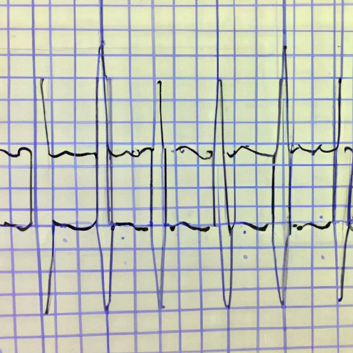
An EKG readout is typically displayed on the monitor as a graph, with each section corresponding to a different phase of the heart’s electrical activity. The readout may also be printed out on a strip of paper or stored digitally for future reference.
Limitations of an EKG
An EKG is a useful tool for diagnosing cardiovascular conditions, but there are some limitations. An EKG cannot detect conditions such as high blood pressure or cholesterol levels. In addition, an EKG may not detect some types of arrhythmias or other abnormalities.
The Role of Electrodes in an EKG: Explaining How the Process Works
The electrodes on the patient’s skin detect small electrical signals produced by the heart. In order for an EKG to accurately measure these signals, the electrodes must be properly placed on the patient’s body.
Placement of Electrodes
The electrodes are typically placed on the patient’s chest, arms, and legs. The exact placement depends on the type of EKG being performed. For instance, a 12-lead EKG requires the electrodes to be placed in specific locations on the patient’s body.
Understanding the Signal Path
Once the electrodes are in place, the electrical signals generated by the heart are detected by the electrodes and sent to the EKG machine. The machine amplifies the signals and displays them on the monitor. The healthcare provider then interprets the results to diagnose any potential problems.
A Look at the Electrical Signals Measured by an EKG
An EKG measures the electrical signals generated by the heart. There are several different types of signals that can be measured by an EKG, including the P wave, the QRS complex, and the T wave.
Different Types of Signals
The P wave is the first signal measured by an EKG and corresponds to the contraction of the atria. The QRS complex is a combination of three signals and corresponds to the contraction of the ventricles. The T wave is the last signal measured by an EKG and corresponds to the relaxation of the ventricles.
Interpreting the Results
The healthcare provider will interpret the results of the EKG to diagnose any potential problems. Abnormalities in the P wave, QRS complex, and T wave can indicate the presence of a cardiovascular condition. For example, an abnormally shaped P wave can indicate atrial fibrillation.

The Benefits and Risks Associated with an EKG
An EKG is a useful tool for diagnosing cardiovascular conditions. However, it is important to understand the potential benefits and risks associated with the procedure.
Advantages of an EKG
An EKG is a non-invasive procedure that can provide valuable information about the functioning of the heart. It is quick and easy to perform, and the results can be used to diagnose and treat various cardiovascular conditions. According to a study published in the Journal of Cardiovascular Nursing, “The EKG is a valuable tool to assess the electrical activity of the heart and can be used to detect and diagnose numerous cardiac conditions.”
Potential Risks of an EKG
Although an EKG is generally considered safe, there are some potential risks associated with the procedure. These risks include skin irritation, allergic reactions, and electric shock. In addition, the results of an EKG may not be accurate if the electrodes are not placed correctly or if the patient moves during the procedure.
A Step-by-Step Guide to Performing an EKG
An EKG is a useful tool for diagnosing cardiovascular conditions. Here is a step-by-step guide to performing an EKG:
Preparing the Patient
Before beginning the procedure, the healthcare provider should explain the procedure to the patient and answer any questions they may have. The patient should be instructed to lie still during the procedure.
Setting Up the EKG Machine
The healthcare provider should then set up the EKG machine and attach the electrodes to the patient’s chest, arms, and legs. The electrodes should be connected to the EKG machine via wires.
Taking the Measurements
The healthcare provider should then take the measurements by pressing the start button on the EKG machine. The machine will amplify the signals and display them on the monitor.
Interpreting the Results
The healthcare provider will then interpret the results of the EKG to diagnose any potential problems. Abnormalities in the P wave, QRS complex, and T wave can indicate the presence of a cardiovascular condition.
Conclusion
An EKG is a useful tool for diagnosing cardiovascular conditions. It is important to understand how an EKG works in order to interpret the results. This article has explored the anatomy and mechanics of an EKG, as well as its benefits and risks. A step-by-step guide to performing an EKG has also been provided.
(Note: Is this article not meeting your expectations? Do you have knowledge or insights to share? Unlock new opportunities and expand your reach by joining our authors team. Click Registration to join us and share your expertise with our readers.)
