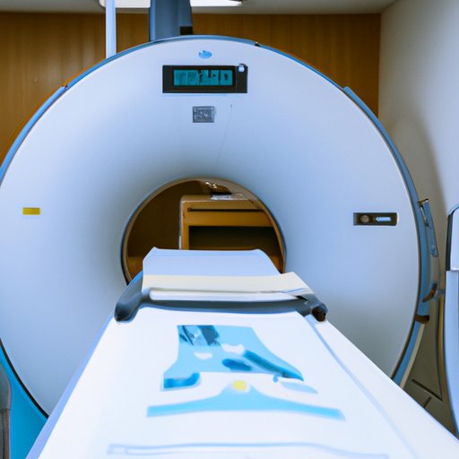
Overview of PET Scan Technology
Positron emission tomography (PET) scans are an advanced type of imaging technology used to identify disease or injury in the body. The scans involve the use of radioactive tracers that help radiologists pinpoint areas of abnormal activity in the body. PET scans are often used to detect cancer, heart and brain conditions, and other illnesses.
Definition of PET Scan
PET scans use a special camera that detects the movement of radioactive material inside the body. The camera produces detailed images that allow doctors to see how the organs and tissues in the body are functioning. This information can help to diagnose or monitor certain diseases and conditions. According to the National Institutes of Health, “PET scans are used to detect and measure levels of different types of metabolic activity in the body.”

How PET Scans are Different from Other Types of Imaging Tests
PET scans are different from other types of imaging tests such as X-rays, CT scans, and MRIs. These tests produce images that reveal the structure of the body, while PET scans provide information about the function of the organs and tissues. PET scans are also able to detect changes in the body at a much earlier stage than other imaging tests.
How PET Scans are Used in Diagnoses and Treatment
PET scans are an invaluable tool for diagnosing and treating a variety of medical conditions. They can help to identify diseases or injuries that may not be visible on other types of imaging tests. They can also be used to monitor the progress of treatments such as chemotherapy or radiation therapy.
Identifying Disease or Injury
PET scans can be used to detect the presence of cancer, heart and brain disorders, and other diseases. They can also be used to determine the extent of a disease or injury, which helps doctors to plan the most effective treatment. According to a study published in the journal Radiology, “PET scans are particularly useful in detecting small lesions and tumors that may not be visible on other imaging tests.”
Monitoring Treatment Progress
PET scans can also be used to monitor the progress of treatments such as chemotherapy and radiation therapy. By monitoring the activity of the tracer, doctors can determine if the treatment is having the desired effect. PET scans can also help to identify any new areas of disease or injury that may have developed since the last scan.
What Happens During a PET Scan
Before undergoing a PET scan, it is important to understand what will happen during the procedure. Knowing what to expect can help to reduce anxiety and ensure that the scan goes as smoothly as possible.
Preparation for the Scan
Before the scan, you will be asked to fast for several hours. You will also need to drink a special solution containing a radioactive tracer. This tracer will travel throughout the body and help to highlight areas where there is abnormal activity.
The Scanning Procedure
During the scan, you will lie on a table and be moved into the scanner. The scanner will take pictures of the body from multiple angles. The entire process usually takes about 30 minutes.
Post-Scan Care
After the scan, you will need to drink plenty of fluids to help flush out the tracer from your body. You should also try to limit your exposure to other people for a few hours after the scan, as the tracer could be passed on through contact.
The Role of the Radiologist in a PET Scan
The results of the PET scan will be interpreted by a radiologist. The radiologist will look at the images produced by the scan and make recommendations based on what they see. Depending on the results, they may suggest further testing or refer you to a specialist.
Interpreting the Results
The radiologist will examine the images produced by the scan and look for any areas of abnormal activity. They will then provide a report detailing their findings and any further tests that may be necessary.
Providing Suggestions for Further Testing
If the radiologist identifies any areas of concern, they may suggest further testing. This could include additional imaging tests such as CT scans or MRIs, or a biopsy to confirm a diagnosis. The radiologist may also provide advice on treatment options.

Understanding the Different Types of Radioactive Tracers Used in PET Scans
There are several different types of radioactive tracers that can be used in PET scans. Each type of tracer has its own advantages and disadvantages. It is important to understand these differences in order to make an informed decision about which tracer is best suited for your individual needs.
Commonly Used Tracers
The most commonly used tracers are fluorodeoxyglucose (FDG), 18F-fluorodeoxyglucose, and 11C-methionine. FDG is the most widely used tracer, as it is highly sensitive and can detect even small areas of abnormal activity. 18F-fluorodeoxyglucose and 11C-methionine are both less sensitive than FDG, but they can still be useful in certain cases.
Advantages and Disadvantages of Each Type
Each type of tracer has its own advantages and disadvantages. FDG is the most sensitive tracer and can detect even small areas of abnormal activity, but it has a short half-life and must be used quickly. 18F-fluorodeoxyglucose has a longer half-life and is less expensive, but it is not as sensitive as FDG. 11C-methionine is also less sensitive than FDG, but it can be used to detect certain types of tumors.

Exploring the Benefits of PET Scans
PET scans offer numerous benefits for patients. They can provide detailed information about the functioning of the organs and tissues, which can help to improve the accuracy of diagnoses. They can also detect diseases and injuries at an early stage, which can help to improve outcomes.
Improved Accuracy of Diagnosis
PET scans can provide more accurate information than other types of imaging tests. Because they measure metabolic activity, they can detect even small areas of abnormal activity, which can help to improve the accuracy of diagnoses. A study published in the journal Nuclear Medicine Communications found that PET scans had a higher accuracy rate than other imaging tests when diagnosing certain types of cancer.
Early Detection of Disease
PET scans can also be used to detect diseases and injuries at an early stage. This can help to improve patient outcomes as early detection often leads to better treatment options. According to a study published in the journal Clinical Cancer Research, PET scans can help to detect cancer at an earlier stage, which can lead to improved survival rates.
Potential Risks Associated with PET Scans
Although PET scans offer many benefits, there are some potential risks associated with the procedure. It is important to understand these risks before undergoing a PET scan.
Radiation Exposure
The most common risk associated with PET scans is radiation exposure. The tracers used in the scan emit low levels of radiation, which can increase the risk of developing certain types of cancer. However, the amount of radiation emitted is usually very small and does not pose a significant health risk.
Unnecessary Testing
PET scans can also lead to unnecessary testing. If the scan reveals an abnormality, further tests may be recommended to confirm the diagnosis. This could lead to additional costs and unnecessary stress for the patient.
False Positive Results
Another potential risk of PET scans is false positive results. This occurs when the scan indicates the presence of a disease or injury, when in fact there is none. False positive results can lead to unnecessary treatments and further testing.
(Note: Is this article not meeting your expectations? Do you have knowledge or insights to share? Unlock new opportunities and expand your reach by joining our authors team. Click Registration to join us and share your expertise with our readers.)
